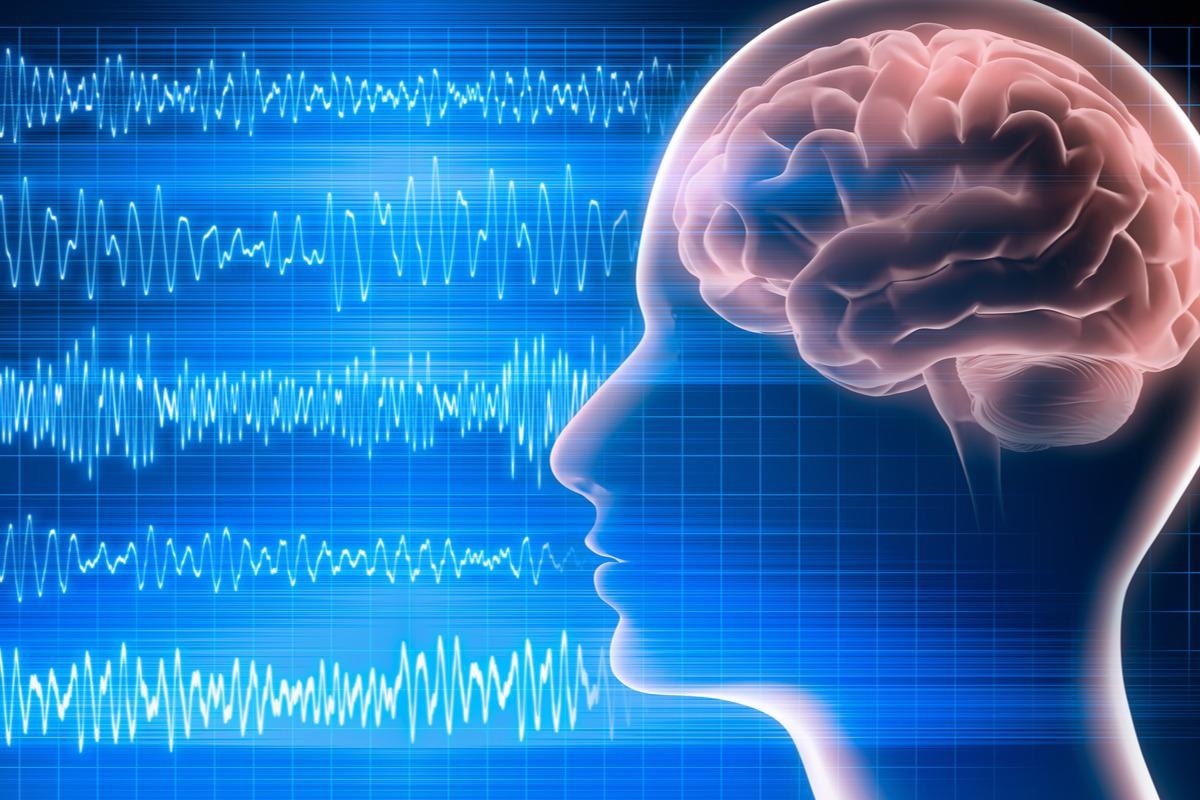
Researchers study EEG patterns in dying man to understand brain activity at the end of life

In a recent Frontiers in Aging Neuroscience study, researchers describe their observations from continuous electroencephalography (EEG) recordings obtained from the brain of a dying 87-year-old man who was experiencing a heart attack after a traumatic subdural hematoma.

Conscious processing in the human brain
Within the healthy human brain, consciousness, memory, and perception activities that occur while individuals are awake, dreaming, and meditating can all be monitored by visualizing brain waves on an EEG. For example, when an individual is conscious, their EEG will typically reflect an increase in thalamocortical activity accompanied by an increase and synchronization in gamma brain waves greater than 35Hz, which are the fastest brain waves produced within the brain.
Comparatively, alpha waves on an EEG, which are the most common type of waves to be observed in a healthy human brain, are often dominant when an individual is processing information, particularly that which is acquired from the visual cortex. When alpha brain waves are dominant, they likely have an inhibitory function on other parts of the brain that are not currently being used.
Whereas delta brain waves are dominant when an individual is trying to complete tasks, theta waves are instead dominant during memory recollection, particularly when the individual recalls verbal and spatial memories.
Taken together, each of the brainwaves is responsible for different neuronal activities that allow for communication, perception, and memory retrieval to be possible. Previous research has found that the same interplay between these brain waves that occur during the recollection of memories in a conscious brain also occurs during memory flashbacks during a near-death experience (NDE).
Previous studies on the neuronal response to death
The conventional belief that neuronal activity declines during NDEs has been challenged over the past several years. For example, rodent studies have provided evidence that the phase-coupling between gamma brainwaves to alpha and theta brainwaves occurs in the first 30 seconds following cardiac arrest and is accompanied by a rise in gamma brainwaves. Similar surges in gamma brainwave activity have also been observed during asphyxia and hypercapnia events.
Aside from animal studies, there have been limited published reports on the neuropsychological processes in the dying human brain. To this end, the current study is the first study to provide continuous EEG recordings from a dying human brain.
About the patient
The current study involved an 87-year-old male patient who presented to the emergency department after falling. Initially, the patient had a Glasgow Coma Scale (GCS) of 15; however, this score rapidly declined to 10. He simultaneously experienced anisocoria, an unequal pupil size, and a bilateral reaction to light.
After confirming that the patient had experienced bilateral acute subdural hematomas (SDH) through computed tomography (CT) scans, a left decompressive craniotomy was performed to remove the hematoma. Although the patient remained stable for two days following this procedure, he continued to decline due to continuous seizures identified through EEG.
Soon after, the patient’s EEG activity demonstrated a burst suppression pattern accompanied by ventricular tachycardia, apneustic respirations, and clinical cardiorespiratory arrest. These findings led the patient’s family to sign a “Do-Not-Resuscitate (DNR),” which ceased any additional treatment until he was allowed to pass away.
EEG findings
Four distinct time points were analyzed by EEG, which included:
- Interictal interval (II) window: 385 to 415 seconds after the clinical seizure was first diagnosed
- Left suppression (LS) window: 510 to 540 seconds between suppression of left and bilateral hemispheric activity
- Bilateral suppression (BS) window: 690 to 720 seconds, wherein the bilateral hemispheric activity ceased, and clinical cardiac arrest occurred
- Post-cardiac arrest (post-CA) window: 810 to 840 seconds between cardiac arrest and the end of EEG recording
Throughout the entire EEG recording, both absolute and relative spectral power exhibited activity levels below 25 Hz. After bilateral hemispheric activity ended, a temporary increase in both absolute narrow- and broad-band gamma brainwaves increased; however, these levels quickly declined after the patient experienced cardiac arrest to levels below all previous time point recordings.
A reduction in delta brainwaves was observed between the II and the LS window, which further declined during the post-CA window. More specifically, delta activity declined by 14.4% between LS and BS time windows at 42% and 27.6%, respectively; however, the post-CA time window experienced a slight increase in delta brainwaves at 37.3%.
After both LS and BS time windows, absolute theta activity decreased and remained stable throughout the post-CA period. Cross-frequency coupling analysis also demonstrated a strong modulation of both low- and broad gamma power occurs due to alpha brainwave activities.
Study takeaways
The EEG recordings from the current study demonstrate that during the transition period to death, the human brain experiences a surge in absolute gamma power, even after the neuronal activity in both hemispheres ceases, that ultimately declines after cardiac arrest.
Furthermore, the researchers observed an intricate interplay between low- and high-frequency brainwaves that occurs after neuronal activity gradually ceases and persists until all blood flow to the brain is cut off due to cardiac arrest.
Several factors may have contributed to the EEG activity observed here, some of which include the lack of oxygen that may have increased cortical excitability after the patient’s injury, as well as the impact of anesthesia on neuronal oscillations. Nevertheless, it appears that the dying human brain experiences varying activity changes during the transition to death.
Vicente, R. et al. (2022) "Enhanced Interplay of Neuronal Coherence and Coupling in the Dying Human Brain", Frontiers in Aging Neuroscience, 14. doi: 10.3389/fnagi.2022.813531. https://www.frontiersin.org/articles/10.3389/fnagi.2022.813531/full#h6
Posted in: Medical Science News | Medical Research News
Tags: Aging, Anisocoria, Blood, Brain, Cardiac Arrest, Coma, Computed Tomography, Cortex, CT, Frequency, Heart, Heart Attack, Neuroscience, Oxygen, Research, Seizure, Tomography

Written by
Benedette Cuffari
After completing her Bachelor of Science in Toxicology with two minors in Spanish and Chemistry in 2016, Benedette continued her studies to complete her Master of Science in Toxicology in May of 2018.During graduate school, Benedette investigated the dermatotoxicity of mechlorethamine and bendamustine; two nitrogen mustard alkylating agents that are used in anticancer therapy.
Source: Read Full Article