A tough attachment between rotator cuff, bone achieved through unique fibrous architecture


Engineers often use nature to inspire new materials and designs. A discovery by a multi-institutional team of researchers and engineers about how tendon and bone attach in the shoulder joint has uncovered previously unsuspected engineering strategies for attaching dissimilar materials. The discovery also sheds new light on how the rotator cuff functions and on why rotator cuff repairs fail so frequently.
Guy Genin, the Harold and Kathleen Faught Professor of Mechanical Engineering in the McKelvey School of Engineering at Washington University in St. Louis, and Stavros Thomopoulos, the Robert E. Carroll and Jane Chace Carroll Professor of Orthopaedic Surgery at Columbia University, led a team that discovered a previously unknown fibrous architecture between the rotator cuff tendons and their bony attachments in the shoulder. Results of the work were published in Science Advances Nov. 26.
Rotator cuff tears—among the most common tendon injuries in adults—occur when tendons pull away from or break near the bone. Thirty percent of adults over age 60 have a tear, and more than 60% of adults over age 80 have a tear. Surgery to repair the tears has a high failure rate, ranging anywhere from 30% to 90% depending on age and other factors. Genin, Thomopoulos and their teams have been studying the mechanobiology of these tissues for several years.
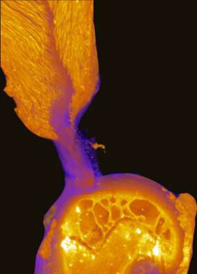
To take a closer look at the enthesis, or the transitional material where each of the four rotator cuff tendons attaches to the bone, the team applied a novel micro computed tomography (microCT) technique. The images revealed a hidden site in the supraspinatus tendon enthesis of mouse shoulders where tendon fibers directly inserted into bone over about 30% of the well-known attachment footprint. Through biomechanical analysis, coupled with numerical simulations, they found that the toughness of the healthy rotator cuff arises from the composition, structure and position of the enthesis as the architecture of the fibrous soft tissues interacts with that of the bone. It was the first time researchers have been able to see both the soft and hard tissues in the rotator cuff simultaneously.
“When [lead author] Mikhail Golman first showed us these images, we realized that much of the old picture of how tendon and bone interact had to be redrawn,” Genin said. “The fiber system there seems like fibers in a rope, and we can understand much about where the toughness comes from by understanding how these fibers break sequentially when they are next to the bone. It’s a new way of thinking about how to attach different materials.”
After the team found the hidden site, there were further discoveries as they progressed.
“Every experiment we did revealed fascinating new features of the attachment system,” Thomopoulos said. “We quickly realized that fundamental aspects of this problem needed to be rethought from scratch. Our goal was to understand where the healthy rotator cuff gets its toughness and strength and under what conditions it ruptures. We found that the toughness of the rotator cuff varies as a function of shoulder position, helping to explain differences in the injury patterns seen in patients.”
The team found that the toughness of the rotator cuff comes from having the fibers help to build the bridge between tissue and bone. Toughness refers to how much energy is required to break a structure, while strength refers to how hard one has to pull to break it, Genin said.
“We found that there’s actually a tradeoff between strength and toughness with these fiber systems,” Genin said. “If you reduce the overall strength by allowing some of the fibers to break, you can actually make the structure tougher because the amount of energy absorbed increases.”
Genin said their results showed that replicating the fiber structure is essential for successful and pain-free healing after rotator cuff repair.
Source: Read Full Article