Neural Mechanisms of Social Anxiety Disorder

Anxiety is an adaptive response that is of great benefit to all life forms. It triggers fear learning, i.e., survival instincts that alert an organism to its surroundings and the dangers that prevail in it. However, extreme anxiety that is inappropriate to a circumstance and is prolonged is a negative emotion, labeled as a social anxiety disorder (SAD). In SAD, negative emotions are stored longer in the memory than others that are less provoking.
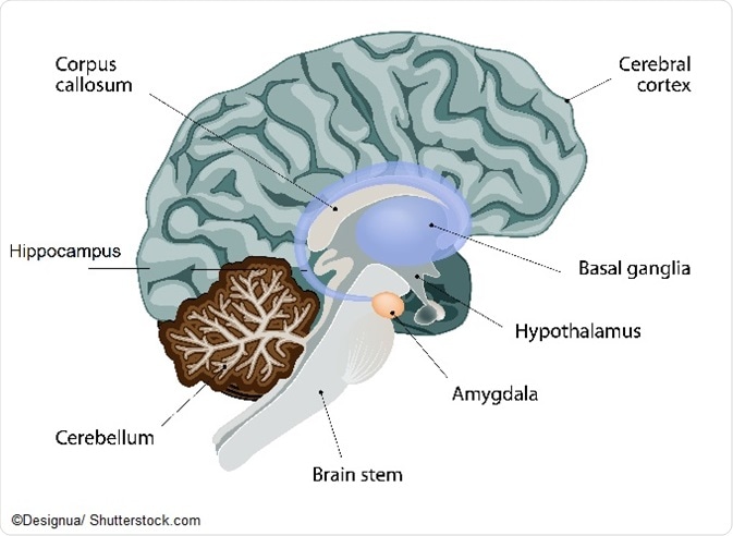
Although the precise neural mechanisms that play a role in anxiety disorders are yet to be fully determined, the limbic system is known to play a vital role in the development and type of emotions we experience and express. The limbic system comprises the:
- Hypothalamus: deals with homeostasis and with the autonomic system;
- Amygdala: the key source that processes emotions, emotional behaviors, and fear conditioning;
- Hippocampus: stores and converts short-term memory into long term.

Till date, consistent hyperactivity of the amygdala and the insula (deep-brain structures) are the known triggers of SAD. Research on humans and other species using functional magnetic resonance imaging (fMRI) has proved that SAD presents with heightened responses to stimuli that may be social or nonsocial.
Place of the amygdala in anxiety
Fear conditioning is acquired and expressed by the amygdala and involves long-term potentiation in the nuclei. The amygdala receives inputs from various parts of the brain and sends them after process to the autonomic and somatosensory centers. Expressions of fear and learning can be stopped by making the amygdala inoperative.
Experiments with rats have shown that when the amygdala of the rat is removed, it totally loses its sense of fear, because it loses its memory of fear. When it sees a cat, it does not fear it because it has no memory of having seen a cat before and been afraid of it. In human experiments, after treatment involving CBT and other medications, persons with SAD show decreased symptoms of amygdala activity.
The activities in the amygdala are regulated by:
- chemicals called neurotransmitters.
- previous mnemonic experiences of fear conditioning and their memory, conveyed by the prefrontal cortex (PFC).
Neurotransmitters that influence SAD
A neurotransmitter is a chemical substance that helps in movement of neural impulses. Any imbalance in these chemicals may lead to a risk of psychiatric disorders.
- Acetylcholine—takes care of learning, memory, voluntary movements, and sleep.
- Dopamine—relates to attention, learning, and movement.
- Serotonin—has a role in mood, appetite, sleep, and kinds of aggressive behavior.
- Norepinephrine—responsible for appetite and alertness.
- Epinephrine—plays a role in glucose metabolism and energy.
- GABA (Gamma-Amino Butyric Acid)—plays a vital role in anxiety and excitation.
Prefrontal cortex
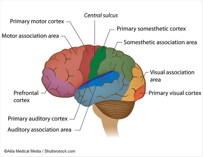
The PFC is the brain region that most differentiates humans from other mammal species, consisting of a specific gene morphology and expression. Located in the frontal lobe, the main role of the PFC is concerned in regulating social and cognitive behavior, decision making, and personality expression. Studies have shown that abnormality in the PFC in anxious individuals is due to a weak connect between the PFC and the amygdala. That is, a higher amygdala activity is related to a lower PFC activity, and the inverse. The PFC uses the baseline level of information from the amygdala and transforms it into the relevant positive or negative emotion.
Many brain areas such as the medial and orbital PFC and the vmPFC, neural substrates, are key in regulating anxiety behaviors. These structures take part in elucidating stimuli from experience, altering behavior patterns based on reward and punishment criteria, and estimating responses to social situations.

Hippocampus
This part of the brain receives signals from the amydgala and processes them in relation to the existing information regarding it. In other words, it deals with episodic memories that can be recalled at will. While the amydgala stores fear memories, the hippocampus forms associations of an emotion with its object. Anxiety disorders that involve fear suggest that threat from the stimuli has not been deleted because of dysfunction in the hippocampus. A decreased hippocampus volume due to damage shows abnormal avoidance learning; this increases vulnerability to avoidance behaviors, which is a risk factor for SAD.
Hypothalamus
While encountering fear or anxiety, the hypothalamic–pituitary–adrenal axis (HPA) gets activated. This axis consists of a set of interactions (among the hypothalamus, pituitary, and adrenal glands) that controls stress reactions. Studies suggest that when stress hormones are continuously released, they impact the levels of serotonin (hormone of well-being) in the HPA. The HPA axis is variously impacted in individuals, depending on the person’s reaction to the stressor or on the stressor itself. Corticosteroids are a class of hormones important for brain development; the HPA axis has a prominent role in the production of this hormone during anxiety moments.
The decade of the 1990s saw immense research undertakings on neurocircuitry mechanisms related to anxiety and fear, which have brought this field closer to improved treatment options for social disorders.
Sources:
- https://www.ncbi.nlm.nih.gov/pmc/articles/PMC4315464/
- https://www.ncbi.nlm.nih.gov/pubmed/19188539
- https://www.ncbi.nlm.nih.gov/pmc/articles/PMC4142809/
- https://www.ncbi.nlm.nih.gov/pmc/articles/PMC4342048/
- https://www.ncbi.nlm.nih.gov/pubmed/11520922
- http://webspace.ship.edu/cgboer/limbicsystem.html
- https://www.ncbi.nlm.nih.gov/pubmed/18019606
- https://canlabweb.colorado.edu/files/understanding_anxiety.pdf
- http://www.umm.edu/health/medical/reports/articles/anxiety-disorders
Further Reading
- All Anxiety Content
- What is Anxiety?
- Anxiety Causes
- Anxiety Symptoms
- Anxiety Diagnosis
Last Updated: May 24, 2019
Source: Read Full Article