new price of viagra in canada
")
The research team led by Professor Hongsoo Choi from DGIST in the Department of Robotics and Mechatronics Engineering has developed a microrobot capable of forming neural networks and sectioning hippocampal tissues in an in vitro environment in an ex vivo state.
Through the joint research with the team led by Dr. Jongcheol Rah from Korea Brain Research Institute, the possibility of analyzing structurally and functionally connected neural networks using a microrobot in an in-vitro environment during cell delivery and transplantation has been confirmed. The work is published in the journal Advanced Materials, and the research findings are expected to be applied in various fields, including neural networks, cell therapy products, and regenerative medicine.
Cell therapy products and cell delivery technology have been developed to regenerate nerve cells damaged by diseases; in recent years, various technologies involving microrobots capable of precise, disulfiram medication interactions minimally invasive cell delivery has been gaining recognition. Previous studies on cell delivery and neural network connections using microrobots only verified structural and functional connections of cells at the cellular level.
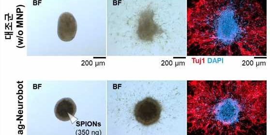
The research team led by Professor Choi used microrobots in which neural network connection can be practically applied. This technology used microrobots to enable the analysis of neural networks functionally connected in an ex vivo environment and cell delivery; the brain tissue of a laboratory mouse was used to conduct the experiment.
The research team first attached superparamagnetic iron oxide nanoparticles to the principal nerve cells of the hippocampus of the laboratory mouse to fabricate the Mag-Neurobot in a three-dimensional spherical form. Magnetic nanoparticles were attached to the outside of the robot so that the robot could move to a desired location by reacting to external magnetic fields. Safety was also verified via a biocompatibility test, in which the magnetism of the robot did not affect the growth of nerve cells.
The research team placed the microrobot in the hippocampus tissue section of the mouse through magnetic field control. Through immunofluorescence staining, the team observed that the cells in the microrobot and the cells in the hippocampus tissue section were structurally connected through neurites.
Furthermore, a microelectrode array (MEA) was used to stimulate the nerve cells in the microrobot to determine whether the nerve cells delivered by the microrobot exhibits typical electrophysiological characteristics. It was verified that electric signals are typically propagated through the nerve cells within the hippocampus tissue section.
Accordingly, the research team confirmed that the nerve cells delivered by the microrobot could functionally form cells and neural networks inside the hippocampus tissue section of a laboratory mouse. In addition, the team demonstrated that the microrobot could perform the roles of delivering nerve cells and forming artificial neural networks.
Dr. Choi of DGIST said, “We have proven that a microrobot and nerve tissues of a mouse brain can be functionally connected through an electrophysiological analysis.”
“The technology developed in this study is expected to be utilized for verifying a precisely targeted treatment in neurological disorder and cell therapy fields.”
More information:
Eunhee Kim et al, A Neurospheroid‐Based Microrobot for Targeted Neural Connections in a Hippocampal Slice, Advanced Materials (2023). DOI: 10.1002/adma.202208747
Journal information:
Advanced Materials
Source: Read Full Article