Protein Crystallography Common Problems, Tips, and Advice

For most proteins that have had their structure determined, this has been done using protein crystallography. When the macromolecular structure of a protein is determined in this way, it is firstly crystalized by supersaturation, which causes each protein molecule to stack with its neighbors in a repeating pattern. The sample then undergoes x-ray crystallography, which is used to examine the structure of various crystal structures.
X-rays generated by the instrument are diffracted by electrons present in the crystal, causing the x-ray photons to be scattered elastically. This means that they possess the same energy both before and after scattering takes place, with only the direction that the photon is traveling in changing. Areas of different electron density scatter the x-ray photons with different intensities, and thus both the positions and bond types of each atom can be determined by the diffraction pattern that is generated.
Skip to:
- Common difficulties faced by protein crystallographers
- Ten tips to consider when creating crystals
- Twinning and other complex issues
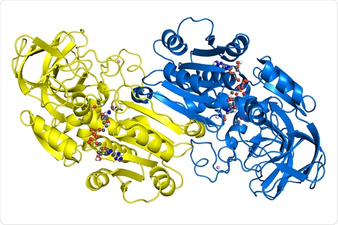
 Crystallographic structure of alcohol dehydrogenase. (petarg | Shutterstock).
Common difficulties faced by protein crystallographers
Most importantly, the crystallized protein samples must be highly pure and stable. In reality, around 50% of the volume of a biomolecular crystal is made up of solvent, which is usually water in the case of proteins, along with other impurities such as small molecules and metal ions.
Other crystals composed of bulk metals or salts do not exhibit this problem, and for this reason, also possess a far larger number of bonds between neighbors than do macromolecular crystals. This makes crystals for protein crystallography highly delicate, sensitive to over-drying when exposed to air, and temperature sensitive. Additionally, they diffract x-rays relatively poorly and become damaged when exposed to radiation, which occurs during x-ray crystallography.
Although the presence of large gaps filled with water between each macromolecule in the crystal is a source of the above problems, it is also advantageous in discovering the true structure of proteins as they would be found in vivo.
Creating crystals suitable for crystallography requires careful manipulation of temperature, pH, and concentration, among other factors.
Ten tips to consider when creating crystals
- The starting protein sample must be pure, often achieved by chromatography
- The protein must be dissolved in the minimum quantity of solvent without forming aggregates
- The proteins must be stable within the solvent
- Supersaturation must be obtained to achieve a suitable crystal
- Proteins should be as close as possible and ordered similarly
- Nucleation begins the crystallization process, and as few as possible should be generated
- Unexpected routes to crystal generation may provide better results, therefore consider experimenting with different methods
- Keep the solution in the optimal conditions during crystallization to ensure they are uniform
- Ensure that the solvent is also pure, and that impurities are not introduced during crystallization
- Once suitable crystals are obtained, keep them safe from temperature extremes, kinetic shock, or other damaging events.
Twinning and other complex issues
Other highly specific problems encountered during protein crystallography will often require a trained and experienced crystallographer to identify and solve. For example, crystals may exhibit twinning due to the growth of two separate crystal lattices from the same point, or highly noticeable favored refraction of x-ray photons from a particular direction. In the latter case, it is best to choose the direction with the highest resolution.
Sources
- Protein crystallography for aspiring crystallographers or how to avoid pitfalls and traps in macromolecular structure determination – ncbi.nlm.nih.gov/pmc/articles/PMC4080831/
- The solvent component of macromolecular crystals – ncbi.nlm.nih.gov/pmc/articles/PMC4427195/
- Introduction to protein crystallization – ncbi.nlm.nih.gov/pmc/articles/PMC3943105/
Further Reading
- All Crystallography Content
- Protein Crystallization
- X-Ray Crystallography
- Protein Structure Determination
- Microseeding Explained
Last Updated: May 23, 2019

Written by
Michael Greenwood
Michael graduated from Manchester Metropolitan University with a B.Sc. in Chemistry in 2014, where he majored in organic, inorganic, physical and analytical chemistry. He is currently completing a Ph.D. on the design and production of gold nanoparticles able to act as multimodal anticancer agents, being both drug delivery platforms and radiation dose enhancers.
Source: Read Full Article