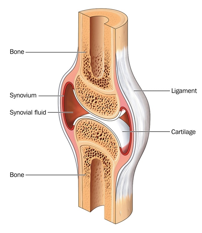
What is Cartilage?

Cartilage is an important structural component of the body. It is a firm tissue but is softer and much more flexible than bone.
Cartilage is a connective tissue found in many areas of the body including:
- Joints between bones e.g. the elbows, knees and ankles
- Ends of the ribs
- Between the vertebrae in the spine
- Ears and nose
- Bronchial tubes or airways
Cartilage is made up of specialized cells called chondrocytes. These chondrocytes produce large amounts of extracellular matrix composed of collagen fibres, proteoglycan, and elastin fibers. There are no blood vessels in cartilage to supply the chondrocytes with nutrients.
Instead, nutrients diffuse through a dense connective tissue surrounding the cartilage (called the perichondrium) and into the core of the cartilage. Due to the lack of blood vessels, cartilage grows and repairs more slowly than other tissues.

Cartilage type
Cartilage is categorized into three types which include:
Hyaline cartilage
This is a low-friction, wear-resistant tissue present within joints that is designed to bear and distribute weight. It is a strong, rubbery, flexible tissue but has a poor regenerative capacity.
Elastic cartilage
Elastic cartilage is more flexible that hyaline cartilage and is present in the ear, larynx and epiglottis.
Fibrocartilage
Fibrocartilage is a tough and inflexible form of cartilage found in the knee and between vertebrae.
Articular cartilage
Articular cartilage is the hyaline cartilage that lies on the surface of bones. This cartilage is often described in terms of four zones between the articular surface and the subchondral bone which include:
The surface or superficial tangential zone
This cartilage covers the articular surface and has a smooth contour that allows gliding of the ends of the bones and resists shear. It forms around 10% to 20% of articular cartilage thickness and has the highest collagen content of all the zones.
The collagen fibrils are densely packed and are aligned in a highly organized manner parallel to the articular surface. The chondrocytes in this zone are elongated in shape.
The middle (or transitional) zone
The middle zone makes up 40% to 60% of the articular cartilage volume. The collagen fibrils are thicker and aligned loosely and not in parallel to the surface. Chondrocytes in this layer are more rounded.
The deep zone
This makes up 30% of the cartilage. The collagen fibrils are large in diameter and aligned perpendicular to the articular surface. This layer has the highest proportion of proteoglycan and lowest concentration of water. The chondrocytes are arranged in a columnar fashion, parallel to the collagen fibers.
The calcified zone
This lies directly on the subchondral bone and contains small cells in a chondroid matrix that has apatitic salts scattered through it.
Sources
- www.med.nyu.edu/…/…sic%20science%20articular%20cart%20and%20OA.pdf
- http://www.cartilagehealth.com/images/artcartbiomech.pdf
- faculties.sbu.ac.ir/…/Cartilage%20[Compatibility%20Mode].pdf
Further Reading
- All Cartilage Content
- Cartilage Diseases
- Cartilage Growth
Last Updated: Feb 26, 2019

Written by
Dr. Ananya Mandal
Dr. Ananya Mandal is a doctor by profession, lecturer by vocation and a medical writer by passion. She specialized in Clinical Pharmacology after her bachelor's (MBBS). For her, health communication is not just writing complicated reviews for professionals but making medical knowledge understandable and available to the general public as well.
Source: Read Full Article