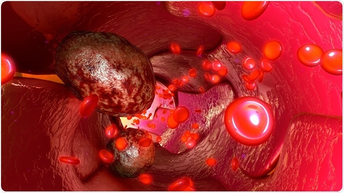
Circulating Tumor Cells and Cancer

Circulating tumor cells (CTCs) are an of population cancer cells that enter the bloodstream after detaching from the primary tumor and propagate to distant tissues to form metastases.

What are CTCs?
Metastasis is a process of developing secondary tumors at different parts of the body that are distant from the primary site of the lesion. Metastatic lesions are more aggressive and account for most of the cancer-related deaths. CTCs play a major role in this process. In order to form metastatic growths, CTCs must pass through certain steps that are crucial for the metastatic process.
Firstly, CTCs must detach from the primary lesion and intravasate into the bloodstream. Intravasation is a process by which CTCs invade through the basal membrane into the blood or lymphatic vessels. Once into the circulation, these CTCs propagate along with numerous other blood cells unless they reach a target tissue. The next important steps are to extravasate into microvessels of the target tissue, adapt to the new microenvironment, and eventually proliferate and colonize to form metastases.
Successful formation of the metastatic growth primarily depends on the ability of CTCs to evade the host’s immune system, adapt to new environment, survive, proliferate, and trigger neoangiogenesis (formation of new blood vessels in tumors). Taken together, it is now clear from many scientific findings that CTCs hold a remarkable promise in understanding in-depth mechanisms involved in the deadly metastatic process.
Detection of CTCs
So far, CTCs have been identified in many cancer types, including breast, colon, lung, prostate, bladder, and gastric cancer. The detection and isolation methods are mainly based on physical or biological properties of CTCs. Physical properties including size and density of the cells and electrical properties of their cell membrane are utilized to differentiate between CTCs and other blood cells. In contrast, methods based on biological properties utilize epithelial cell adhesion molecules (EpCAM) present on the cell surface to positively enrich CTCs in blood samples of patients with cancer.
Another approach is the use of CD45 antibody-coated magnetic beads that remove most of the leucocytes and negatively enrich CTCs in blood samples. Recently, a new method has been developed that does not involve positive or negative enrichment. This method involves direct lysis of blood sample to remove red blood cells and subsequent fluorescence microscopic analysis of residual nucleated cells that are bound onto substrates and stained with fluorescence-labeled antibodies. The presence of CTCs in the blood can give a chance to get samples of cancer tissues through liquid biopsy, which is less invasive.
Problems Related to CTC Detection
Since most of the CTC detection methods rely on epithelial markers, CTCs that undergo epithelial-to-mesenchymal transition and, thus, do not express epithelial makers escape the detection process. Another limitation is that the number of detectable CTCs in blood is generally very less, which imposes an extra burden in getting an accurate estimation. Moreover, most of the methods involve too many steps and harsh sample preparation process, which can significantly affect cell viability.
Importance of CTCs in Cancer Therapy
Due to their significant involvement in the metastatic process, CTCs are considered as a potential biomarker for evaluating prognosis in patients with metastatic lesions. They have also become potential therapeutic targets for controlling or preventing tumor metastasis. Some of the important applications of CTCs in cancer patients include analyzing the response of cancer patients to a particular therapy, categorizing high-risk and low-risk patients by detecting CTCs in early-stage cancers, identifying patients based on the need for surgery and/or chemotherapy, and monitoring the recurrence of the tumor during chemotherapy.
Analysis of the molecular properties of CTCs is another important aspect that can provide significant insight into genotypic and phenotypic characteristics of a tumor without performing a biopsy. In addition, since detection of CTC does not require any invasive method, it is relatively easy to frequently monitor tumor progression and check incidences of drug-resistance through molecular profiling of CTCs.
CTC as a Prognostic Biomarker
In recent years, many studies have been performed to detect the numbers of CTCs in blood to evaluate cancer prognosis and outcomes. It has been found in one of such studies that breast cancer patients with CTCs ≥ 5 have shorter median progression-free and overall survival compared to patients with CTCs less than 5.
A similar scenario has also been observed in patients with metastatic prostate cancer. In the case of colorectal cancer patients, CTC number ≥ 3 is found to be associated with significantly worse median progression-free and overall survival compared to CTC number less than 3.
Sources
- http://www.annclinlabsci.org/content/43/3/295.full
- https://www.ncbi.nlm.nih.gov/pmc/articles/PMC3126032/
- https://www.ncbi.nlm.nih.gov/pmc/articles/PMC5453029/
- http://science.sciencemag.org/content/341/6151/1186
Further Reading
- All Circulating Tumor Cell Content
- Microfluidic Enrichment in Single-Cell Analysis
- Circulating Tumor Cells and Cancer
Last Updated: Oct 9, 2018

Written by
Dr. Sanchari Sinha Dutta
Dr. Sanchari Sinha Dutta is a science communicator who believes in spreading the power of science in every corner of the world. She has a Bachelor of Science (B.Sc.) degree and a Master's of Science (M.Sc.) in biology and human physiology. Following her Master's degree, Sanchari went on to study a Ph.D. in human physiology. She has authored more than 10 original research articles, all of which have been published in world renowned international journals.
Source: Read Full Article