generico viagra sandoz

The rapid spread of the severe acute respiratory syndrome coronavirus 2 (SARS-CoV-2) resulted in the coronavirus disease 2019 (COVID-19) pandemic. Although most individuals infected with SARS-CoV-2 are asymptomatic or experience mild symptoms, some succumb to severe infection with chronic lung injury and acute respiratory distress syndrome (ARDS).

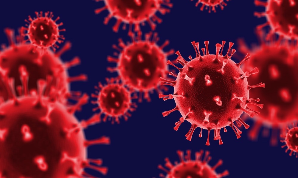
Study: SARS-CoV-2 Spike triggers barrier dysfunction and vascular leak via integrins and TGF-β signaling. Image Credit: Billion Photos / Shutterstock.com
Background
Lung pathology reports of severely infected patients indicated the occurrence of edema stemming from epithelial and endothelial barrier dysfunction. Although previous studies found that this condition is triggered by a hyperinflammatory response, the exact inducing factor for epithelial/endothelial hyperpermeability remains unclear.
SARS-CoV-2 belongs to the Coronaviridae family and possesses a positive-sense ribonucleic acid (RNA) genome that encodes four structural proteins, of which include the spike (S), membrane (M), nucleocapsid (N), and envelope (E), and non-structural proteins.
Antibodies eBook

The SARS-CoV-2 S glycoprotein, which is present in the outer coating of the virus, binds with angiotensin-converting enzyme 2 (ACE2) receptors of the host cell and establishes infection. The S protein consists of two domains including S1 and S2.
S1 contains the receptor-binding domain (RBD) that binds with ACE2, while S2 promotes the fusion of the virus-host cell membranes. Cathepsin L, Furin-like proteases, and transmembrane protease, serine 2 (TMPRSS2) are other crucial host factors for SARS-CoV-2 infection.
In addition to binding to ACE2, the S glycoprotein is also associated with various other cell-surface factors, such as integrins and heparan sulfate-containing proteoglycans (HSPG). These factors predominantly promote SARS-CoV-2 entry into the host cell. The association of the viral S protein with these factors has been linked with signaling pathways that contribute to lung pathology.
During the infection of SARS-CoV-2, S1 can be shed from the surface of the virions after binding to the ACE2 receptor. This suggests that shed-S1 could also interact with endothelial and epithelial cells; however, the mechanisms behind this interaction are not fully understood. Furthermore, the host factors involved in these interactions have not been identified.
Previous studies have established how viral proteins like flavivirus non-structural protein 1 (NS1) interact with endothelial cells. This interaction induces signaling cascades that subsequently promote the disruption of cellular structures that are essential for endothelial barrier integrity associated with the endothelial glycocalyx layer (EGL) and intercellular junctional complexes.
About the study
A recent Nature Communications study analyzed whether the SARS-CoV-2 S protein influences endothelial and epithelial barrier dysfunction in vitro and vascular leak in vivo.
The authors hypothesized that the local concentration of the S protein accumulated in capillaries within tissues would be higher than the levels in patients’ sera. Therefore, the concentration of S used in this study was similar to circulating levels in severely infected COVID-19 patients.
The concentration of S used for the experiments ranged from 2.5 µg/mL to 20 µg/mL. However, most experiments were conducted at 10 µg/mL.
Key findings
The in vitro and in vivo experimental findings indicated that virion-associated full-length S, soluble trimeric S, and recombinant RBD could trigger barrier dysfunction. The first and second mechanisms by which SARS-CoV-2 induces barrier dysfunction is due to its interactions with ACE2-negative non-permissive cells and during the infection of virus-permissive cells.
The third mechanism responsible for this barrier dysfunction is through the shedding of soluble S1 after enzymatic cleavage following ACE2 interactions on a cell. The expression of S on the surface of infected cells that can interact with nearby cells may also contribute to this phenomenon.
The authors conjectured the role of S-mediated barrier dysfunction in COVID-19 pathogenesis, which is the spread of SARS-CoV-2 from the lungs to the blood and then into distal organs, the latter of which is where virus-complacent cells reside. This conjecture was validated through in vivo experiments using a mouse model.
To this end, clinical samples of COVID-19 patients sufficiently facilitated barrier dysfunction. Thus, in addition to its role in viral entry to the host cell, the S protein also interacts with glycosaminoglycans (GAGs) and integrins to induce vascular leakage through activation of the transforming growth factor beta (TGF-β) pathway.
Transcriptional analyses demonstrated that S glycoprotein regulates the expression of transcripts involved in modulating the extracellular matrix (ECM). Experimental analysis elucidated the underlying mechanisms, wherein TGF-β, GAGs, and integrins were associated with barrier dysfunction.
In vivo experiments similarly demonstrated that the SARS-CoV-2 S glycoprotein triggers vascular leak in the lungs of mice, which was reversed by integrins.
Conclusions
Taken together, the current study provided the mechanistic explanation for the overproduction of TGF-β during COVID-19, which has been correlated with disease severity. Furthermore, full-length S and RBD of SARS-CoV-2 can independently mediate barrier dysfunction and vascular leak.
In the future, more research must be conducted to understand the structural basis of the mechanisms.
- Biering, S. B., Gomes de Sousa, F. T., Tjang, L. V., et al. (2022) SARS-CoV-2 Spike triggers barrier dysfunction and vascular leak via integrins and TGF-β signaling. Nature Communications 13(7630). doi:10.1038/s41467-022-34910-5
Posted in: Medical Science News | Medical Research News | Disease/Infection News
Tags: ACE2, Acute Respiratory Distress Syndrome, Angiotensin, Angiotensin-Converting Enzyme 2, Blood, Capillaries, Cell, Chronic, Coronavirus, Coronavirus Disease COVID-19, covid-19, Edema, Enzyme, Genome, Glycoprotein, Growth Factor, in vitro, in vivo, Lungs, Membrane, Mouse Model, Pandemic, Pathology, Protein, Receptor, Research, Respiratory, Ribonucleic Acid, RNA, SARS, SARS-CoV-2, Serine, Severe Acute Respiratory, Severe Acute Respiratory Syndrome, Spike Protein, Structural Protein, Syndrome, Vascular, Virus

Written by
Dr. Priyom Bose
Priyom holds a Ph.D. in Plant Biology and Biotechnology from the University of Madras, India. She is an active researcher and an experienced science writer. Priyom has also co-authored several original research articles that have been published in reputed peer-reviewed journals. She is also an avid reader and an amateur photographer.
Source: Read Full Article