buy online synthroid supreme suppliers without prescription


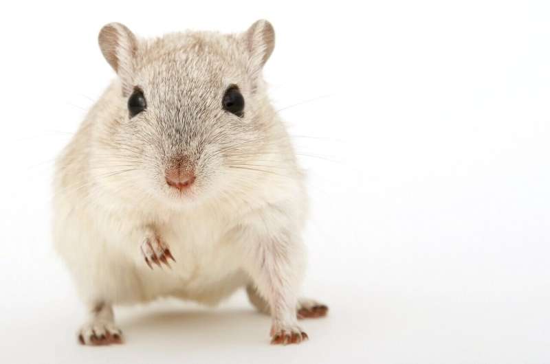
Baby mice might be small, but they’re tough, too. For their first seven days of life, they have the special ability to regenerate damaged heart tissue. Humans, on the other hand, aren’t so lucky: any heart injuries we suffer could lead to permanent damage. But what if we could learn to repair our hearts, alli sims middle name just like baby mice?
A team of researchers led by UNSW Sydney have developed a microchip that can help scientists study the regenerative potential of mice heart cells. This microchip—which combines microengineering with biomedicine—could help pave the way for new regenerative heart medicine research.
The study is featured on the cover of today’s issue of the journal Small.
“We’ve developed a simple, reliable, cheap and fast way to identify and separate these important mouse heart cells,” says lead author Dr. Hossein Tavassoli, a biomedical engineer and stem cell researcher at UNSW Medicine & Health who conducted this work as part of his doctoral thesis.
“Our method uses a microchip that’s easy to fabricate and can be made in any laboratory in the world.”
The process for identifying and separating mice heart cells is rather complex.
First, scientists need to separate the right kind of heart cells (called proliferative cardiomyocytes) from other types of cells present in the heart.
Their next challenge is keeping the cells alive.
“Newborn mice heart cells (called proliferative cardiomyocytes) are very sensitive,” says Dr. Vashe Chandrakanthan, a senior research fellow at UNSW Medicine & Health and co-senior author of the study.
“Only about 20 per cent usually survive the conventional isolation and separation process. If we want to study these cells, we need to isolate them before they undergo cell death.”
Dr. Tavassoli says that this new method is much more efficient.
“We reduced the stress applied on these cells by minimising the isolation and processing time,” he says. “Our method can purify millions of cells in less than 10 minutes.
“Almost all of the cells survived when we used our microfluidic chip—over 90 per cent.”
The spiral-shaped device is a microfluidic chip—that is, a chip designed to handle liquids on tiny scale. It filters cells according to their size, separating the cardiomyocytes from other cells. The chip costs less than $500 to produce, making it cheaper than other isolation and separation methods.
This tool will make it easier for researchers to study how baby mice repair their hearts—and whether humans might be able to use the same technique.
“Heart disease is the number one killer in the world,” says Dr. Tavassoli. “In Australia, someone dies of heart disease every 12 minutes, and every four hours a baby is born with a heart defect.
“We hope that our device will help accelerate heart disease research.”
Characterising mice heart cells
Once the heart cells were separated from other cells with the help of their chip, the researchers seized the opportunity to study the cells’ physico-mechanical properties—that is, the way they respond to force.
This involved asking questions like ‘How do these individual heart cells beat?’, ‘Do the cells have distinct features?’ and ‘What are their differences in size, shape and elasticity?’.
The findings could provide new insights for developing materials that repair heart tissue, like cardiac patches, scaffolds and hydrogels.
“The fast, large-scale characterisation of cells’ physico-mechanical features is a relatively new field of research,” says Dr. Tavassoli, who originally trained as an engineer before specialising in medicine.
“This is the first time microfluidic technology has been used to study mechanical properties of baby mouse heart cells.”
A multipurpose microchip
Dr. Chandrakanthan says that even though the microchip was created for baby mouse heart cells, it could potentially be adapted for use in other types of cell applications.
“The principles are compatible with isolating cardiomyocytes from mouse heart cells of all ages,” he says.
“We could potentially also use this method to separate not only the heart cells, but all sorts of cells from different organs.”
Dr. Tavassoli says this method could also help other areas of medical research, including cardiac biology, drug discovery and nanoengineering. He is currently conducting research at the Garvan Institute and Lowy Cancer Research Centre on how this method could help cancer diagnosis.
Source: Read Full Article