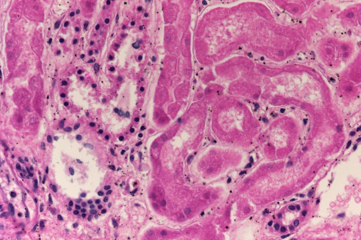
Impact of acute kidney injury associated gene expression in human kidney cells

Acute kidney injury (AKI) is a condition that is associated with high morbidity and mortality and is common in critically ill patients. It has been reported that more than 10% of hospitalized patients and 50 % of critically ill patients in intensive care units develop this condition. Currently, the therapy for AKI involves providing supportive care and kidney replacement in case of severe kidney failure. Therapies to prevent AKI and to promote recovery do not still exist.
 Study: Transcriptomic responses of the human kidney to acute injury at single cell resolution. Image Credit: Arisang/Shutterstock
There is a lack of comprehensive knowledge on the cellular mechanisms that are associated with AKI and the response to injury provided by kidney cells. A high incidence of AKI has been observed in coronavirus disease 2019 (COVID-19) patients.
Arecent study released as a preprint on the bioRxiv* server attempts to perform a comparative single-cell census of human kidneys employing samples from individuals suffering from AKI and controls without the condition. The findings from the study may enhance the knowledge on AKI and help design effective treatment strategies to prevent or treat the condition.
How was the study performed?
The present study was conducted on kidney tissue samples obtained from eight individuals with severe AKI. Of the eight individuals in the study four were diagnosed with COVID-19. Samples were collected from individuals who had succumbed to complications due to respiratory infections. The control samples were collected from individuals without AKI either after nephrectomy or post-mortem.
Single-cell transcriptome census of human AKI was performed using single-nuclei RNA sequencing (snRNA-seq) of kidney samples from individuals with AKI and normal controls. Findings from principal component analysis (PCA) suggest that AKI was the main determinant of the cell type-specific and global gene expression changes that were observed when the samples from controls and individuals with AKI were compared. Further, differences in gene expression exist even amongst individuals with AKI indicating the existence of distinct molecular subtypes in AKI.
The response of specific kidney cell types to AKI was assessed by performing differential gene expression analysis using DESeq2.
It was found that significant transcriptomic responses to AKI were observed in the kidney cells in the proximal tubule (PT), the thick ascending limb of the loop of Henle (TAL), the distal convoluted tubule (DCT), and connecting tubules (CNT), cells types belonging to the cortex and outer medulla regions of the kidney which is at higher risk of ischemic and hypoxic injuries.
The differentially expressed genes that were identified were those that encode markers of kidney injury including neutrophil gelatinase-associated lipocalin/lipocalin 2 (LCN2), kidney injury molecule 1 (HAVCR1), and insulin-like growth factor-binding protein 7 (IGFBP7).
The kidney cell sources that express these markers in response to injury were also identified. As reported earlier, CNT and collecting duct principal cells (CD-PC) were the major sites where LCN2 was upregulated and in the case of HAVCR1 it was PT. Further, Secreted phosphoprotein 1 (SPP1) which encodes osteopontin was upregulated in non-leukocyte kidney cell types. In correlation with previous reports, IGFBP7 mRNA was upregulated in cells of the PT, and additionally, it was also upregulated in podocytes and TALs.
The pathways to which the differentially expressed genes belong were identified and it was found that some of the genes that are upregulated in AKI were related to the inflammatory response-associated pathways such as tumor necrosis factor-alpha, interferon-gamma, and interleukin signaling, pathways for hypoxia response, and epithelial to mesenchymal transition.
Further, there was simultaneous upregulation of functional pathways in all the kidney tubule cell types in response to AKI indicating a common response exhibited across all the cell types. Similar to earlier reports the present study found that genes associated with molecule transport and metabolism were downregulated in response to AKI.
Gene expression changes specific to kidney cell types in individuals with AKI in the presence or absence of COVID-19 were compared. It was found that the transcriptomic responses to AKI in kidney cells of COVID-19 affected individuals were not significantly different when compared to samples from individuals suffering from AKI due to other causes.
Novel injury-related kidney cell states were enriched in response to AKI
Subclusterings of the kidney cell types were performed and cells were segregated based on their transcriptomes resulting in the identification of 74 kidney cell populations. These included cell populations that were identified based on known cellular subtypes and additionally, novel cell populations designated as "New" cell populations were identified that represented injury-related cell states.
Further analysis was performed to identify the cell subpopulations that were depleted or enriched in individuals with AKI. The PT is known to be susceptible to injury and it has been reported to have a propensity towards dedifferentiation in AKI. The findings from the present study showed that cells belonging to the PT were significantly depleted aligning with the earlier observations. It was additionally found that medullary TAL, DCT, CNT cells were also depleted in response to AKI. Notably, it was observed that the “New” cell subpopulations belonging to these cell types were enriched in AKI suggesting that the “New” cell subpopulations are injury-associated cell states.
Endothelial cells, interstitial cells, and leukocytes in kidneys which are non-epithelial cell types were neither enriched nor depleted in response to AKI. An exception was one subtype of endothelial cells which was enriched in AKI.
The “New” cell population was further characterized and four “New” cell clusters (PT-New-1-4) associated with PT were identified. It was observed that 31.8% of PT cells in samples from individuals with AKI belonged to one of these four clusters. The marker genes were identified and pathway analysis was performed for PT-New-1-4. Some of the enriched genes and pathways identified were oxidative stress signaling and the nuclear transcription factor erythroid 2-related factor 2 (NRF2) pathway in PT-New -1, the hypoxia response pathway in PT-New 2, the interferon-gamma response, and genes encoding for ribosomal proteins in PT-New 3 and genes associated with epithelial-mesenchymal transition (EMT) in PT-New 4.
The PT-New1-4 were compared with PT-derived injured cell states in mice with renal ischemia-reperfusion injury identified from an earlier study. Four cell states were identified “injured PT S1/S2 cells”, “injured PT S3 cells”, “severe injured PT cells” and “failed repair” cells. It was found that PT-New-1 showed similar characteristics as “injured PT S1/S2 cells”, the PT-New-2 were similar to “injured PT S3 cells”, PT-New 3 and PT-New 4 were similar to the “failed repair” cells.
The individual and combined abundances of PT-New-1-4 were found to be significantly high in samples from individuals with AKI. The distribution of PT-New-1-4 among AKI-affected individuals was found to be heterogeneous e.g. The abundance of PT-New 4 in individuals with AKI was found to vary by a factor of three.
RNAscope in situ hybridizations were performed to identify transcripts overexpressed in PT-New 1-4 clusters. Interleukin-18 (IL18) mRNA was overexpressed in a small number of cells in PT-New 2 and PT-New 4, insulin-like growth factor-binding protein 7 (IGFBP7) was expressed in a substantial number of PT-New 1-4 cells, Interferon-induced transmembrane protein 3 (IFITM3) was expressed in cells in PT-New 1, 3, and 4.
The study additionally found that though the injury responses in kidney epithelial cells are cell-type specific and exhibit inter-individual heterogeneity, they are still associated with common pathways and marker genes.
Conclusion
The findings from the present study reveal the cell type-specific transcriptomic responses in AKI. The study identified novel injury-associated cell states in the tubular epithelium of proximal tubules, thick ascending limbs, and distal convoluted tubules. Four AKI-associated injured cell states were also identified whose transcriptomes corresponded with oxidative stress, hypoxia, interferon response, and epithelial-to-mesenchymal transition. Inter-individual heterogeneity was observed between individuals with AKI which was attributed to cell type-specific abundance of the four injury-related cell states identified. Further AKI-associated changes in the transcriptome were similar both in the presence and absence of Severe acute respiratory syndrome coronavirus 2 (SARS-CoV-2) infection. Based on the findings from this study, single-cell transcriptomics may serve as tools for developing suitable therapies for AKI. Moreover, personalized molecular disease assessment in AKI may also promote the development of personalized therapies to treat this condition.
*Important notice
bioRxiv publishes preliminary scientific reports that are not peer-reviewed and, therefore, should not be regarded as conclusive, guide clinical practice/health-related behavior, or treated as established information.
- Hinze, C. et al. (2021). Transcriptomic responses of the human kidney to acute injury at single cell resolution. bioRxiv. doi: https://doi.org/10.1101/2021.12.15.472619 https://www.biorxiv.org/content/10.1101/2021.12.15.472619v1
Posted in: Medical Research News | Medical Condition News | Disease/Infection News
Tags: Acute Kidney Injury, Cell, Coronavirus, Coronavirus Disease COVID-19, Cortex, Gene, Gene Expression, Genes, Growth Factor, Hypoxia, Insulin, Intensive Care, Interferon, Interferon-gamma, Interleukin, Kidney, Kidney Failure, Leukocyte, Metabolism, Molecule, Mortality, Necrosis, Nephrectomy, Osteopontin, Oxidative Stress, Protein, Respiratory, RNA, RNA Sequencing, SARS, SARS-CoV-2, Severe Acute Respiratory, Severe Acute Respiratory Syndrome, Stress, Syndrome, Transcription, Transcriptomics, Tumor, Tumor Necrosis Factor

Written by
Dr. Maheswari Rajasekaran
Maheswari started her science career with an undergraduate degree in Pharmacy and later went on to complete a master’s degree in Biotechnology in India. She then pursued a Ph.D. at the University of Arkansas for Medical Sciences in the USA.
Source: Read Full Article