Benefits of Multiplex Immunoassays

How do you multiplex an immunoassay?
There are two main ways of multiplexing an immunoassay; by separating the individual assays into different spaces (spatial separation) or by immobilizing antibodies onto different beads based on what they target.
 Jarun Ontakrai | Shutterstock
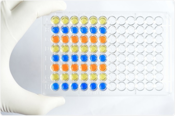
Spatial separation is typically achieved by placing antibodies in different parts of a shared solid surface. In this way, the reaction is only run once on the shared surface, but different areas of the surface would detect different proteins.
This can be used both to detect the presence of multiple proteins, as well as to quantify the amounts of these proteins present in a sample. In this case, a secondary antibody will be used, and the amount of these which have bound to the target analyte can be quantified.
Immobilization of the various antibodies on to individual beads adds the complication of not knowing what antibody is on which beads once mixed. To solve this problem, various dyes can be added to the beads in order to facilitate the separation and identification of beads within this mixture.
Once the analyte has had the chance to bind to these capture antibodies, secondary antibodies can be used to see which beads have captured a protein, and then after analyzing what color these beads are the types of proteins present in a sample can be determined.
Why use a multiplex assay?
The main advantage of a multiplex assay, of course, is that you can analyze multiple proteins simultaneously rather than having to run multiple assays to look for each protein separately.
Running multiple assays means that there could be subtle variations in each run, which may impact the results. Running one assay prevents this variation from having a significant effect so that any difference seen in the levels of various proteins is more likely to reflect the true abundances within the sample.
Another disadvantage of running multiple single assays is this will require more sample. This may not be a problem if the sample is easy to obtain, however, some biological samples are not typically available on mass, thus, reducing the amount of sample needed for assays is a huge advantage. In this instance, multiplex assays can be very beneficial as it is possible to detect and quantify multiple proteins from a smaller sample volume.
How do multiplex immunoassays work in practice?
A study by Ray and co. used a multiplex immunoassay to look for five cytokines in human blood. Cytokines are molecules that are involved in many biological processes, including inflammation and cell differentiation, and the levels of these cytokines present in blood plasma can vary as part of a disease process.
Sepsis is one such disease that is characterized by a change in the levels of cytokines that promote inflammation (pro-inflammatory cytokines), such as IL-1β, TNFα, IL-6, and IL-8. Cytokine levels can also be changed in response to certain compounds, some of which are being investigated as potential therapeutic drugs. Therefore, it is beneficial to have a system where the levels of multiple cytokines can be analyzed at once; this is what Ray and co. intended to do. Here, they developed an assay which could identify levels of IL-1β, TNFα, IL-6, IL-8, and IL-10.
Using a bead-based method, each bead had capture antibodies for the above cytokines and were distinguished by the ratio of dye added. The authors were able to detect concentration of these cytokines while using a limited amount of patient blood sample, and at a reduced cost.
Sources
- www.abcam.com/…/multiplex-immunoassay-techniques-review-of-methods-western-blot-elisa-microarray-and-luminex
- https://www.sciencedirect.com/science/article/pii/S0731708504002559
Further Reading
- All Immunoassays Content
- What are Lateral Flow Assays?
- How do Lateral Flow Immunoassays Work?
- Tyramide Signal Amplification (TSA) Methodology
- Tyramide Signal Amplification (TSA) Applications
Last Updated: Apr 5, 2019

Written by
Dr. Maho Yokoyama
Dr. Maho Yokoyama is a researcher and science writer. She was awarded her Ph.D. from the University of Bath, UK, following a thesis in the field of Microbiology, where she applied functional genomics toStaphylococcus aureus . During her doctoral studies, Maho collaborated with other academics on several papers and even published some of her own work in peer-reviewed scientific journals. She also presented her work at academic conferences around the world.
Source: Read Full Article