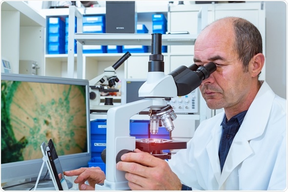
Applications in Light Microscopy

Light microscopy has a number of applications in different sectors including in gemmology, metallurgy and chemistry. In terms of biology, it is one of the least invasive techniques for looking at living cells.

The light microscope can be used to provide information about the activity of cells and to look at very small structures such as nanostructures.
Different adaptations can help to enhance images, such as phase contrast microscopy, which provides contrast between cells and the solution they are in. High resolution 3D imaging can also be used to observe organisms over a period of time.
Fluorescence microscopy is also a good technique for observing specimens that fluoresce and emit light of a different colour. The number of fluorescent proteins has increased, expanding the kinds of sample types that can be looked at, from single molecules to whole organisms. In addition, unwanted side effects have been reduced. However, one limitation of fluorescence microscopy is the overlap of fluorophores, which can make analysis more difficult.
Molecular imaging
Microscopy can be used to explore the time- and space-related dynamics of molecules. Localizing single molecules such as RNA and proteins provides insights into how cells and tissues are organized at the molecular level.
Cell imaging
High content screening (HCS) uses microscopy to identify and study substances in cells such as peptides, RNA and small molecules. Automated microscopes can be used to look at thousands of compounds or genetic alterations and the effects these have. Information about the structure, heterogeneity, kinetics and more can be obtained from the resulting images.
HCS can be used by biotechnology and pharmaceutical companies to screen for potential drug candidates. It allows researchers to consider and rule out a number of different molecules in a short period of time.
This method can also be used to look at genes to find out more about the genome and potentially identify sequences that alter cell phenotype and lead to different diseases.
The process allows for fast analysis of the genome and can identify molecules that have effects on the majority of the 21,000 gene products found in cells.
References
- Cell Communication and Signalling Journal, Light microscopy applications in systems biology: http://www.ncbi.nlm.nih.gov/pmc/articles/PMC3627909/
- Wartburg College Biology department: https://www.wartburg.edu/biology/fluorescentmicro/applications.html
- Wikipedia on high throughput biology: https://en.wikipedia.org/wiki/High_throughput_biology
Further Reading
- All Microscopy Content
- Advances in Fluorescence Microscopy
- Electron Microscopy: An Overview
- Brief History of Microscopy
- Bright field Versus Dark-field TEM
Last Updated: May 29, 2019

Written by
Deborah Fields
Deborah holds a B.Sc. degree in Chemistry from the University of Birmingham and a Postgraduate Diploma in Journalism qualification from Cardiff University. She enjoys writing about the latest innovations. Previously she has worked as an editor of scientific patent information, an education journalist and in communications for innovative healthcare, pharmaceutical and technology organisations. She also loves books and has run a book group for several years. Her enjoyment of fiction extends to writing her own stories for pleasure.
Source: Read Full Article